Risks During Pregnancy
If you have a high-risk pregnancy, you or your baby might be at increased risk of health problems before, during or after delivery. Typically, special monitoring or care throughout pregnancy is needed. Here’s what you can expect

Prenatal diagnostics encompasses all the examinations that are carried out to detect hereditary diseases or physical problems in the unborn child. These procedures are particularly relevant to women with certain risk factors that run in the family.
TESTING FOR GENETIC OR CHROMOSOMAL DISORDERS
A screening test is conducted in mothers who may be at risk. Based on the results of this test, the doctor will make a decision as to whether further tests should be carried out. Today, most chromosome disorders can be established with the so-called ‘First Trimester Test,’ the optimum period for this test being between the 11th and the 14th week of pregnancy. This test takes into account the mother’s age, the results of a maternal blood analysis and the thickness of the unborn child’s neck fold, which is measured by an ultrasound.
ULTRASOUND SCAN
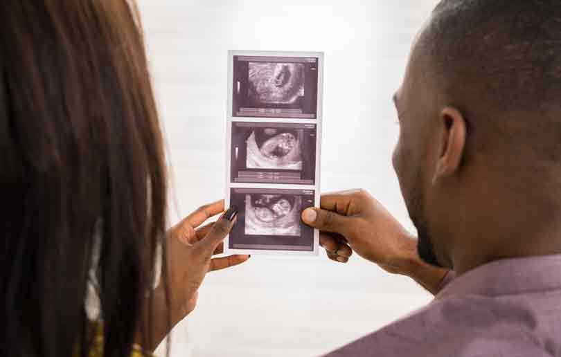
Ultrasound examinations are routinely conducted before pregnancy, to check fertility or cyclic changes in the uterus and ovaries. It is also used once the pregnancy is established, to check on the health of the pregnancy and the growing embryo. The doctor precisely measures the embryo, in order to determine the length of the pregnancy and the date of delivery. The scanner also makes it possible to recognise any abnormalities early on, by measuring the neck fold.
The first ultrasound test takes place between the 10th and 14th week of pregnancy, and the second between the 20th and the 23rd week. There are no known risks for the child in the ultrasound testing. The doctor can usually recognise arms and legs, as well as the heartbeat and any movement of the embryo, early on in the pregnancy. The best time to examine the baby’s organs is in the 20th – 23rd week, at which point, the organs are large enough to permit a detailed sonographic examination and it is possible to clearly see the baby’s face.
CONSIDERATIONS IN THE FIRST TRIMESTER
• The heart is beating visibly
• Position of the foetus in the uterus
• Stage of pregnancy reached
• Multiple or single pregnancy
• Possible chromosome disorders (e.g. Downs Syndrome)
CONSIDERATIONS IN THE SECOND AND THIRD TRIMESTER
• Quantity of amniotic fluid
• Extent of foetal growth
• Evidence of abnormalities
• Position of the placenta
• Doppler examination, in the case of high risk pregnancies (blood circulation in the placenta and foetus)
• 3D ultrasound
In the UAE, only two ultrasound examinations are covered by health insurance. There would need to be a medical reason to justify any further scans.
MEASURING THE NECK FOLD
Between the 10th and 14th week of pregnancy, the skin fold in the neck of the child is measured using ultrasound imaging. The thicker the fold, the larger the risk for Downs Syndrome. This simple method has 80% rate of identifying babies that are affected. A thickened neck fold can also be due to harmless causes. This is why the testing of the neck fold may be combined with an examination of the mother’s blood, which is called ‘serum screening’.
MATERNAL SERUM SCREENING (BLOOD TEST)
The blood test can be carried out between 14 weeks and 20 weeks, plus 6 days to screen for neural tubedefects such as spina bifida (abnormal development of part of the spine and spinal cord) and anencephaly (severely abnormal development of the brain). If you are having the second trimester blood test for Downs Syndrome, neural tube defect screening can be done at the same time. If the test shows that your baby has an increased chance of having a neural tube defect, an ultrasound will be done straight away. A detailed ultrasound scan of the baby when you are around 18 – 20 weeks pregnant can detect almost all babies with a neural tube defect (95%).
If the results of your screening tests indicate the possibility of any abnormalities, a more precise testing method can be used, such as Chorionic Villus Sampling (CVS) or amniocentesis.
FACTORS THAT MIGHT CONTRIBUTE TO A HIGH-RISK PREGNANCY
• Maternal age: Pregnancy risks are higher for mothers older than age 35.
• Lifestyle choices: Smoking, drinking alcohol and using drugs can put a pregnancy at risk.
• Medical history: A history of diabetes, heart disorders, breathing problems, infections, and blood-clotting disorders, such as deep vein thrombosis, can increase pregnancy risks.
• Surgical history: A history of surgery on your uterus, including multiple C-sections, multiple abdominal surgeries or surgery for uterine tumours(fibroids), can increase pregnancy risks.
• Multiple pregnancy: Pregnancy risks are higher for women carrying twins or higher order multiples.


Comments